Distribution and survival of primary sarcoma in Korea: A single center analysis of 2017 cases
Article information
Abstract
Purpose
Distribution and survival of sarcoma in Korea are not well described, after the changing of sarcoma classification on 2013. The researchers investigated the distribution and survival in single center 2017 cases of sarcoma.
Methods
Patients with primary sarcoma, who underwent surgery, were investigated. All cases were collected during a 20 year period (1995–2015) from Samsung Medical Center in Korea. Histopathologic types were classified by World Health Organization (WHO) classification (2013). And overall survival rates were analyzed.
Results
Between 1995 and 2015, 2017 patients were collected. The most frequent type of sarcoma was gastrointestinal tumor (15%), followed by liposarcoma (12%), leiomyosarcoma (9%), dermatofibrosarcoma (6%), giant cell sarcoma (6%). The most common primary site of sarcoma was the intra-abdominal area (45%, including visceral area). Extremities accounted for 26% of all cases. Sixteen percent of sarcoma were located in retroperitoneal area. The overall survival rate was 70.4% (median follow-up time, 36.8 months; range, 0.1–261.3 months). The best prognosis was dermatofibrosarcoma (100%, 5-year survival rate). The worst prognosis was angiosarcoma (39.3%). Survival analysis by the primary site demonstrated favor prognosis in extremities than head & neck, chest lesion.
Conclusion
The researchers reported Korean sarcoma characteristics with using the new WHO classification.
INTRODUCTION
Sarcomas were mesenchymal originated tumors, arising in connective tissue. These were a very rare incidence of overall human malignant tumors, accounting for less than 1% of all new cancer diagnoses [1]. This heterogeneous malignancy tumor consists of 70 different histologic subtypes [2].
Histopathologic classification of sarcoma has changed considerably since 1978 [3]. The World Health Organization (WHO) published new sarcoma classification, representing updated consensus, in 2013. According to this, gastrointestinal tumor (GIST) has included in sarcoma and many cases of malignant fibrous histiocytoma (MFH) were classified as specific sarcoma [4]. Many previous reports about the epidemiology of sarcoma were not based on new classification. Only a few studies described the characteristics of sarcoma, and prognosis with new classification.
In Korea, one study reported epidemiology and prognosis of sarcoma. It was based on a previous classification with low volume of data [5]. Therefore, the aim of the study is to analyze the distribution, characteristics, prognosis of sarcoma based on new classification with a retrospective review of single center 2,017 cases.
METHODS
Data were retrospectively collected from a database of Samsung Medical Hospital, Seoul, Korea, between January 1995 and December 2015. Patients who underwent surgery for primary sarcoma were investigated. Data collected included age, gender, date of surgery, histological type, primary site, completeness of resection, 5-year survival, and overall survival (OS).
Primary sarcoma was defined as a tumor that had not been treated previously, and no evidence of metastasis. Tumors which underwent second surgery due to positive cancer cell on resection margin on first surgery at other hospital were included in this study.
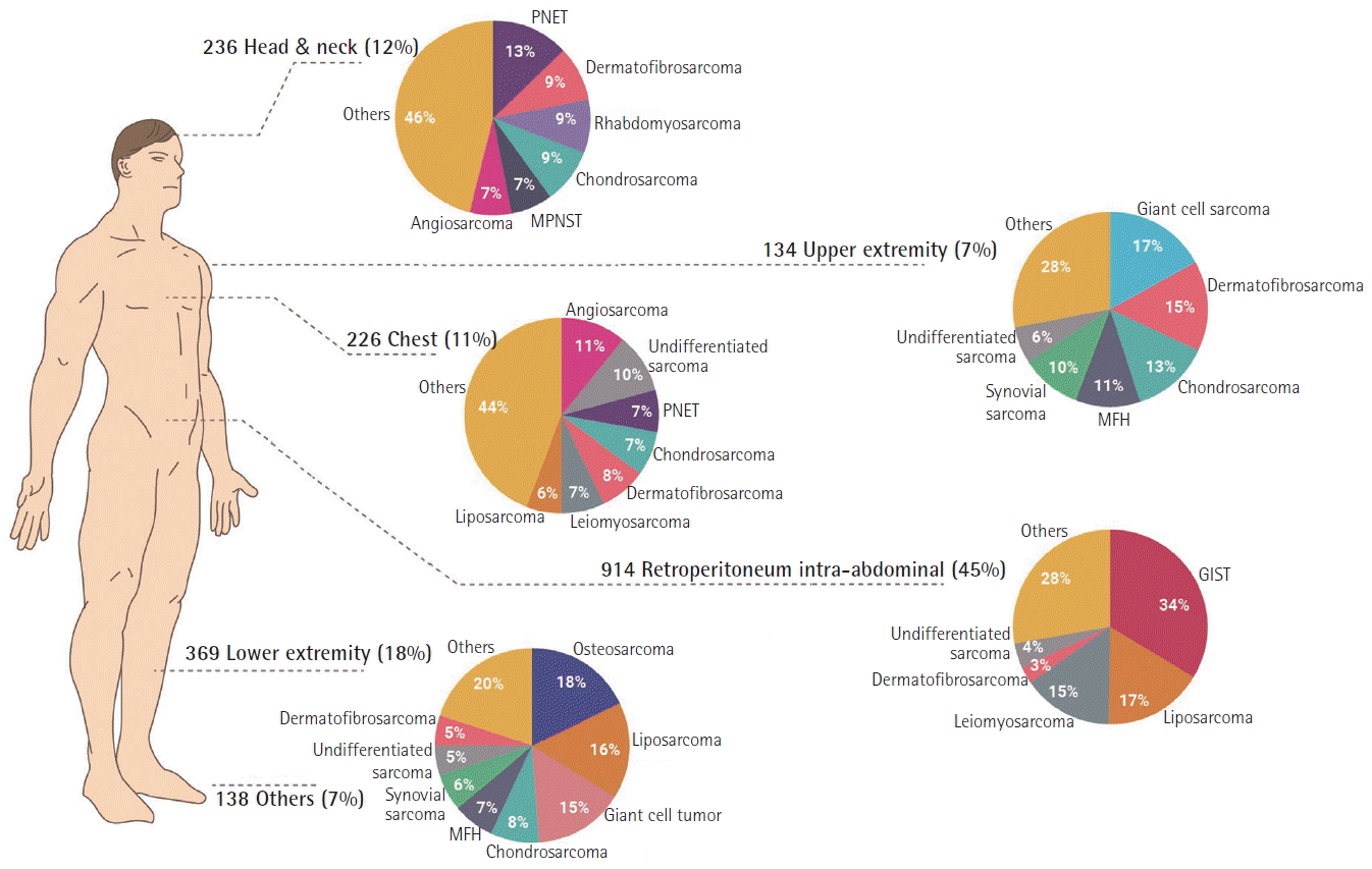
Histologic types were reviewed according to the WHO classification [4]. In the GIST, both high risk group (National Institutes of Health consensus criteria, 2002) and malignant types were included. Tumors were classified according to 15 histologic groups: GIST, liposarcoma, leiomyosarcoma, dermatofibrosarcoma, giant cell tumor, chondrosarcoma, undifferentiated sarcoma, osteosarcoma, MFH, primitive neuroectodermal tumor (PNET), rhabdomyosarcoma, angiosarcoma, malignant peripheral nerve sheath tumor (MPNST), synovial sarcoma, others (solitary fibrous tumor, myxosarcoma, fibrosarcoma, epithelioid sarcoma, clear cell sarcoma, alveolar soft-part sarcoma, and so on). Liposarcomas were subdivided into five subtypes: well-differentiated, myxoid, dedifferentiated, pleomorphic, not otherwise specified (NOS). The following cases were excluded, for the reasons explained in parentheses: mesothelioma (mesothelial tumors of the pleura), neuroblastoma (tumors of the autonomic nervous system), mesenchymomas (an entity not clearly defined), carcinosarcoma (tumors that is mixture of carcinoma), ganglioneuroblastoma (tumors of the autonomic nervous system). Primary sites were divided into six groups: head & neck, chest, abdominal cavity, upper extremities, lower extremities, others (genital organ, pelvic bone, and vascular lesion). The abdominal cavity was subdivided into three groups: intra-abdominal, retroperitoneal, abdominal wall. The researchers illustrated primary sites and proportion of histological subtypes on one figure (Fig. 1).

Subtype distribution according to histologic types, primary sites. PNET, primitive neuroectodermal tumor; MPNST, malignant peripheral nerve sheath tumor; MFH, malignant fibrous histiocytoma; GIST, gastrointestinal tumor.
Completeness of resection was categorized as clear (R0), microscopically positive (R1), grossly positive (R2). Gross resection status was determined with operating record documented by an operating surgeon. Margin status was investigated based on a pathologic report. A clear margin indicated there was no tumor on resection margin microscopically. A microscopically positive margin indicated microscopically extension of cancer cell regardless of distance from the tumor margin. In addition, cases of undetermined margin status in pathologic report with grossly total resection were considered as R1 resection.
Overall survivorships were calculated from the date of surgery to the last known date of follow-up or the date of death. Date of death was identified from the linkage of Korean Statistical Information Service. Disease specific death was not investigated. Based on 15 histologic subtypes of sarcomas and six primary sites, survival rates were calculated for each types and sites. Estimation of survival was calculated by the Kaplan-Meier method and survivorship analysis, being assessed by the log-rank test.
Survival graphs were illustrated and analyzed by grouping similar origin of sarcomas together. Rhabdomyosarcoma, chondrosarcoma, osteosarcoma, synovial sarcoma and leiomyosarcoma were grouped in musculoskeletal group PNET, MPNST and angiosarcoma were in neurovascular group. The other subtypes were grouped in unclassified group including GIST and liposarcoma. On the other hand, survival graphs by primary sites in all kinds of sarcoma were illustrated all together. All analysis was done using SPSS version 23.0 (IBM Corp., Armonk, NY, USA). Statistical significance was defined as P< 0.05.
RESULTS
Subtype distribution
Two thousand, one hundred and fifty-eight patients, who underwent surgery for primary sarcoma, were investigated. One hundred and forty-one patients were excluded because of palliative surgery. The median age was 48 years (range, 0–91 years; mean, 45.6 years). The ratio of male to female was 1.04:1. The seventy-one percent (n= 1,425) of patients underwent grossly complete excision, whereas 157 patients (8%) had grossly margin positive. Among all patients, the most common histologic type was GIST (15%), followed by liposarcoma (12%), leiomyosarcoma (9%), dermatofibrosarcoma (6%), giant cell sarcoma (6%) (Table 1).
The most common primary site was the abdominal cavity (45%), followed by the lower extremity (18%), head & neck (12%), chest (11%), others (7%), and upper extremity (7%). Even when dividing the abdominal cavity into three regions: intra-abdominal (27%), retroperitoneal (16%), abdominal wall (2%), the intra-abdominal was still the most common site of sarcoma. In the head & neck region, PNET (13%) was the most common histologic type. Five types of sarcoma (dermatofibrosarcoma [9%], rhabdomyosarcoma [9%], chondrosarcoma [9%], MPNST [7%], angiosarcoma [7%]) accounted for half of the rest. For the thoracic region (chest), the most common histologies were angiosarcoma (11%), undifferentiated sarcoma (10%), followed by dermatofibrosarcoma (8%), primitive neuroectodermal sarcoma (7%), chondrosarcoma (7%), leiomyosarcoma (7%), and liposarcoma (6%). In the abdominal cavity, GIST (34%) was the most common histologic type and liposarcoma (17%) was the second most common type. For extremity sarcoma, the giant cell tumor was the most common subtype on the upper extremity (17%) but be on the third place on lower extremity (15%). Osteosarcoma (18%), liposarcoma (16%) were the most common types on the lower extremity (Fig. 1).
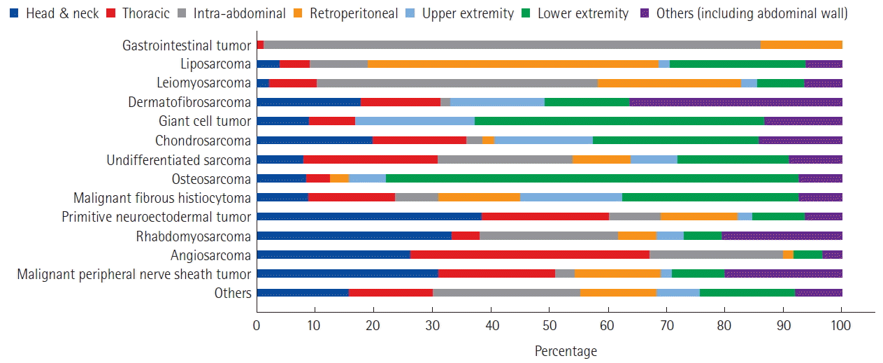
The overall primary site distribution of each histologic subtypes was displayed on Fig. 2. Most of GIST were in abdominal cavity (85%). Retroperitoneal area was accounted for 13%. Only 1% of GIST were in thoracic region. All GIST cases of thoracic region were occurred on esophagus. In liposarcoma, majority of lesion were in retroperitoneal area (50%). Lower extremity was accounted for 23%. The head & neck was the most common primary site of PNET (38%).
Survival
The overall sarcoma survival rate was 70.4%, with a median follow-up time of 36.8 months (range, 0.1–261.3 months). The best prognosis subtype was the dermatofibrosarcoma (100%) and followed by GIST (90%). Whereas angiosarcoma (39.3%) demonstrated the worst prognosis.
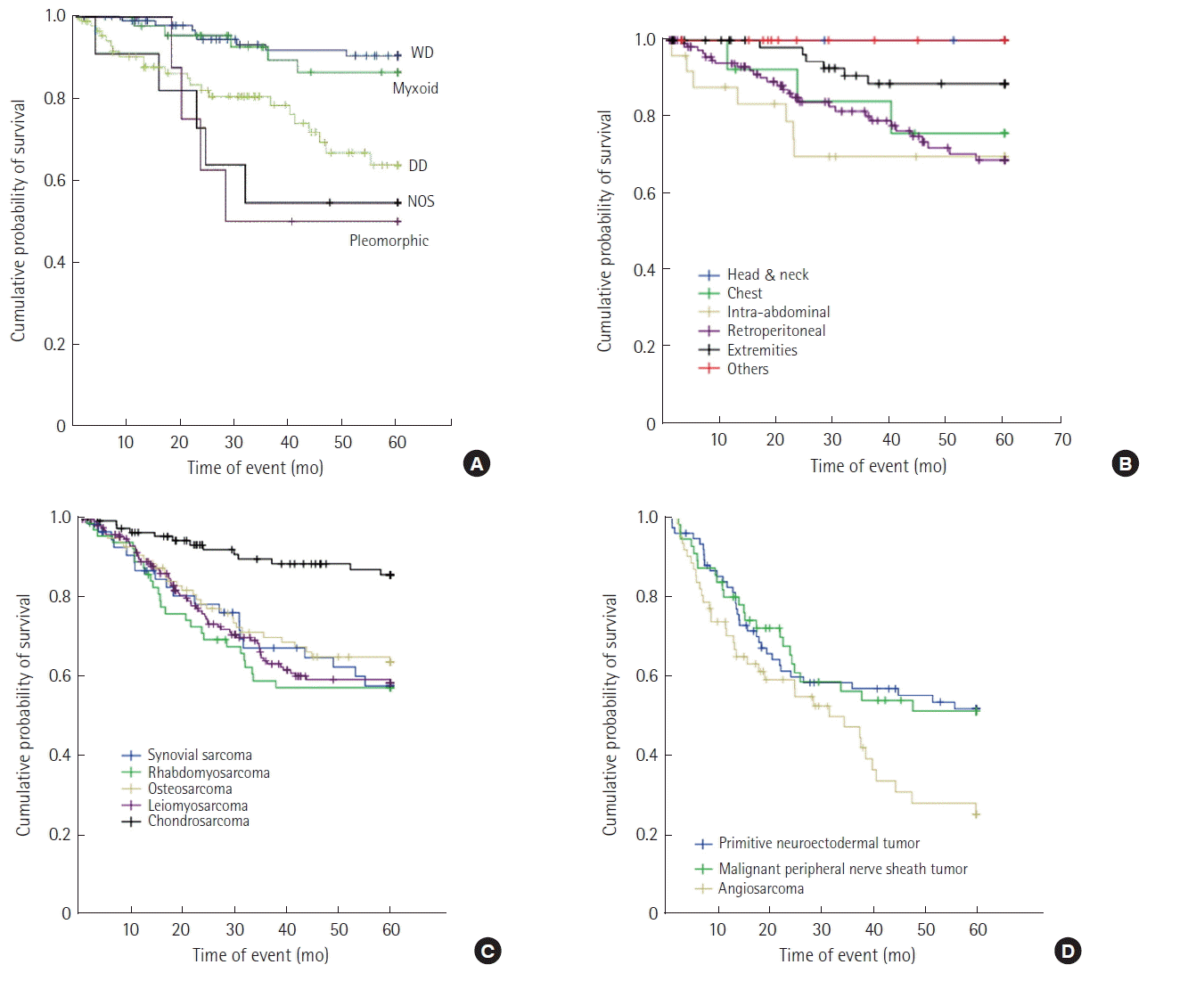
In the liposarcoma group, survivorship was differed depending on subtypes, ranging from 54.5% (pleomorphic) to 91.8% (well-differentiated). When survivorships were compared each subtype with liposarcoma, NOS, well-differentiated liposarcoma (93.5%, P< 0.0001) and myxoid liposarcoma (88.6%, P< 0.009) demonstrated a better survivorship than liposarcoma NOS (55.6%). The 5-year survivorships of each subtype are demonstrated in Fig. 3A. Survivorship comparison by primary sites was higher in extremities (90.5%) than retroperitoneal (77.6%, P< 0.009), intra-peritoneal (72.0%, P< 0.015) (Fig. 3B).

Survival outcome according to subtypes. (A) Five-year overall survival (OS) of liposarcoma for each subtypes, (B) 5-year OS of liposarcoma for each primary site, (C) 5-year OS of musculoskeletal group, (D) 5-year OS of neurovascular group. WD, well differentiated; DD, dedifferentiated; NOS, not otherwise specified.
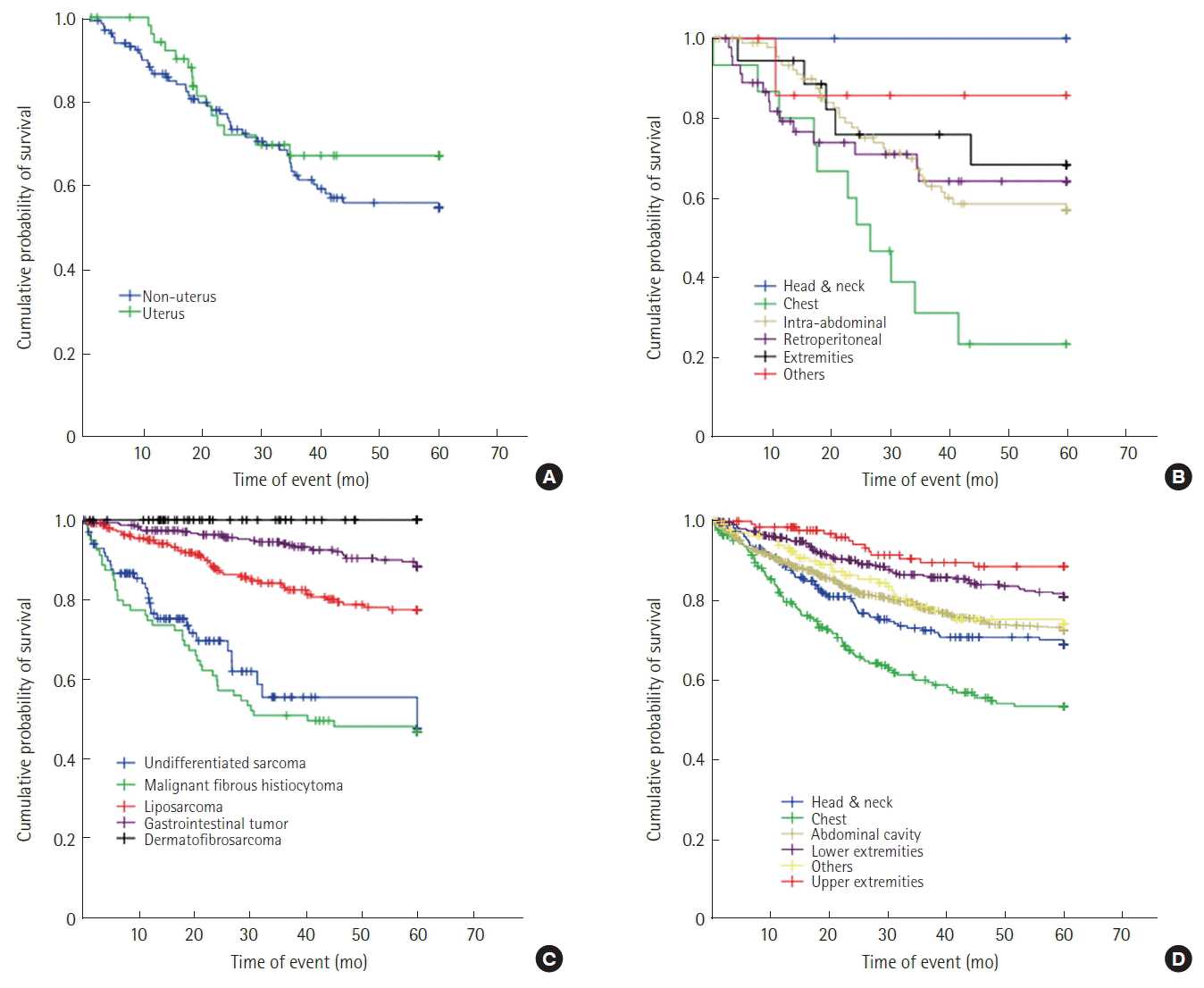
In the musculoskeletal group, chondrosarcoma (87.7%) demonstrated a better prognosis than the other four subtypes (synovial sarcoma [63.6%], rhabdomyosarcoma [58.7%], osteosarcoma [66.3%], leiomyosarcoma [65.2%], P< 0.001). Except chondrosarcoma, the other subtypes were demonstrated a similar progressive decrease in survivorship graph (Fig. 3C). For the neuromuscular group, angiosarcoma (39.3%) had a poor prognosis in 5-year survival than MPNST (56.4%, P< 0.019), PNET (54.7%, P< 0.037). All the three types were demonstrated similar survival graph until 30 months. However, after 30 months of follow-up, angiosarcoma demonstrated a more significant decrease (Fig. 3D). Especially in leiomyosarcoma, uterus group (72.2%) was demonstrated favor survivorship than non-uterus group (62.4), however it was statistically not significant (P< 0.254) (Fig. 4A, B).

Survival outcome according to subtypes. (A) Five-year overall survival (OS) of leiomyosarcoma in uterus, non-uterus group, (B) 5-year OS of leiomyosarcoma for each primary site, (C) 5-year OS of others group, (D) 5-year OS of primary site.
In others group, there was no death in dermatofibrosarcoma patients during 5-year follow-up period. On the comparison with liposarcoma (82.2%) histologic subtype, undifferentiated sarcoma (69.0%), MFH (46.3%) were shown poor prognosis, whereas dermatofibrosarcoma (100%), GIST (90.0%) were better (P< 0.001) (Fig. 4C).
In the comparison of survival rates according to primary sites, the upper extremities (90.2%), and the lower extremities (83.7%) demonstrated less progressive decrease in survivorship graphs than others (79%). Abdominal cavity (76.2%) and head & neck (72.9%) were shown similar curves with others group, but chest group (59.9%) were shown more progressive decrease in survivorship graph. However only upper extremities (P< 0.004), chest region (P< 0.001) were statistically significant (Fig. 4D).
DISCUSSION
The aim of this study was to estimate histologic distribution, distribution of location, survival rates of sarcoma in the Korea. In addition, this is the first study to show a distribution of sarcoma, including GIST in Korea. In the data, the most common histologic type was GIST, accounting 15% of all. Hui previously reported liposarcoma was the most common [6]. This difference was due to the fact that our institute had only an abdominal sarcoma center without an emphasis on limb sarcoma. In addition, the high incidence of GIST in Korea (16–22 per million) might have affected the underestimation of liposarcoma [7,8]. GIST, liposarcoma and leiomyosarcoma were the most common histologic subtypes. This finding was concurrent with previous other reports [5,9,10]. In addition, the most common place of sarcoma was the abdominal cavity, which was more than sum of upper and lower extremities. Previous reports were reported the extremities as the common place. This difference was due to change of sarcoma classification, especially including GIST [4,5,11]. Distribution of histologic types in other lesions (head & neck, thoracic, extremities) was not concurred with any previous study [5,6,12]. It is not clear why the research data demonstrated a different distribution, especially a low proportion of MFH. However, the exception of metastatic cases and changes in diagnostic criteria of MFH might be an influence in result [3]. Distribution of primary site by the histologic types was concurred with other reports [6,8,13]. For instance, most of GIST occurred in the stomach and small intestine. The retroperitoneum and lower extremities were the most common place in liposarcoma. One third of PNET and rhabdomyosarcoma were diagnosed on the head & neck.
In the present study, 5-year OS of GIST (90%) was demonstrated higher than previous reports (50%–65%) [14,15]. It is unclear why our study demonstrated higher survivorship because this study did not investigate characteristics (mitotic index, size) of GIST. Therefore, further research of this findings will be needed.
Regarding to histologic distribution of liposarcoma in this study, well differentiated and de-differentiated type were the most common types. This is concurrent with previous report of Oh et al. [16]. Which analyzed 213 patients’ survival for liposarcoma regarding to histologic subtypes and primary sites. In addition, the 5-year OS according to histologic types was also similar with this research data, favorable 5-year OS in well-differentiated liposarcoma than dedifferentiated liposarcoma and pleomorphic liposarcoma. Previous studies classified inguinal/genitalia as a new group of primary sites in liposarcoma, whereas this study classified inguinal/genitalia, and head & neck as other group altogether. The reason for this classification was that the most of head & neck liposarcoma were occurred in scalp, cheek, skin of neck. Therefore, in this study, the others group demonstrated better survival than retroperitoneal group without death in 5-years.
Chondrosarcoma was found to demonstrate a significant higher 5-year OS (P <0.001) than other subtypes in musculoskeletal group (synovial sarcoma, rhabdomyosarcoma, osteosarcoma, leiomyosarcoma). Stiller et al. [17] also reported the favor prognosis of chondrosarcoma (76.7%) than osteosarcoma (53.9%) [18]. However, previous reports analyzed survivorship by classifying sarcoma as soft tissue group (leiomyosarcoma, rhabdomyosarcoma, synovial sarcoma) and bone sarcoma group (osteosarcoma, chondrosarcoma) [17–19]. There was no recent report of survivorship analysis between chondrosarcoma and rhabdomyosarcoma, leiomyosarcoma.
Leiomyosarcoma demonstrated 65.2% in 5-year OS in present study, which is similar with other reports (64.2%–65.7%) [20,21]. In subgroup analysis between uterus group (72.2%) with non-uterus group (62.4%) in leiomyosarcoma, demonstrated favor survivorship in uterus group than non-uterus group without statistical significance in present study. Lamm et al. [22] that analyzed 95 patients of leiomyosarcoma (50 non-uterus leiomyosarcoma, 45 uterus leiomyosarcoma) also reported the better prognosis in uterus group than non-uterus group (82.6% vs. 41.2%, P< 0.006).
The most of study did not analyze soft tissue sarcoma and bone sarcoma together because of its different subtypes, different natural history and different response to therapy [16–18]. However, this study was conducted distribution and survivorships analysis together. Therefore, It will be need more specific survivorship analysis in future study by subgrouping as soft tissue sarcoma, bone tissue sarcoma, adult (over 18 age) sarcoma, child sarcoma (under 18 age) etc.
There are some limitations in this study. First, it was a retrospective analysis. Second, a pathologic slide review was not conducted. Therefore, there can be misdiagnosed cases due to changing classifications. In addition, as mentioned above, because of characteristics in the study institution (having only abdominal sarcoma center without Limb sarcoma center). Selection bias also exists.
These data demonstrated subtypes and survival rate for sarcoma patients in South Korea by new classification of WHO (2013). The distribution of sarcoma seems to be similar with pervious report. Whereas survival rates were higher than previous reports especially in GIST. The reason of this finding was not clarified; further investigation is needed to confirm this finding.
Notes
CONFLICT OF INTEREST
No potential conflict of interest relevant to this article was reported.

