INTRODUCTION
Gastric cancer is one of the most common cancers and the second leading cause of cancer related deaths worldwide [1,2]. Until now, many studies have investigated the prognostic factors of gastric cancer. For example, tumor size, invasion depth, lymph node involvement and distant metastasis status are known as important prognostic factors [3-5]. Specifically, these factors were sometimes related with operative plans; for instance, the extent of surgery [6,7].
In recent days, signal transduction related growth factor and its receptors were considered to have an important role in carcinogenesis [8-11]. Among them, the human proto-oncogene human epidermal growth factor receptor 2 protein (HER-2, also called c-erbB2 or Her-2/neu) processes tyrosine kinase activity as an encoding signal transduction pathway related to the regulation and differentiation of cell growth [12,13]. In fact, HER-2 oncogene was usually studied in relation to breast cancer, as a poor prognosis factor [9,14]. However, HER-2 oncogene has also appeared as an important prognostic factor in gastric cancer [4,5,8,12,15-21]. Especially, HER-2 oncogene status is related with the depth of invasion, lymph node and distant metastasis of gastric cancer [4,18,19].
Recent targeting therapy of gastric cancer has been gradually applied by using the humanized monoclonal antibody ‘Trastuzumab (Herceptin)’ targeting HER-2 receptor. In this regard, HER-2 oncogene status is becoming important as a biomarker for evaluation and treatment of gastric cancer [8,20,22,23].
The aims of this study were to clarify the association between HER-2 status and patient’s clinicopathologic factors in patients who underwent curative intent gastrectomy.
METHODS
Patients
From June 2011 to May 2015, curative intent gastrectomy was performed on 441 patients. Among them, HER-2 status was tested in 113 patients who were diagnosed with advanced gastric cancer.
Data on clinicopathologic parameters such as age, gender, histological subtype, endoscopic Lauren classification, tumor location, size, presence of lymphovascular invasion, invasion depth, pathologic stage, HER-2 overexpression, recurrence, and survival were obtained from the medical records of the patients. The tumors were staged according to the 7th edition of the American Joint Committee on Cancer staging system [24]. Clinical outcome was evaluated from the date of surgery to the date of the last follow-up day.
Samples and immunohistochemistry
Gastric cancer tissue samples from patients who underwent curative intent gastrectomy were formalin-fixed and paraffin-embedded. These samples (n= 113) were obtained and arrayed manually until April, 2012. Later, it was performed by using an automatic tissue-arraying instrument (BenchMark, Ventana Medical System Inc.). Samples were divided on 3-um tissue sections and then section forms were made neutral-buffered, formalin-fixed, and paraffin-embedded. Tissue sections were fixed using coated slides (Menzel-Glaser: Superfrost plus) and then deparaffinization and hydration was done. Endogenous activity was controlled by incubation with 3% hydrogen peroxide. Antigen retrieval was done, and the primary antibody used (anti-Her2/neu (4B5) Rabbit Monoclonal Primary Antibody). The chromogen used was diaminobenzidine, and the samples were counterstained with hematoxylin.
Interpretation
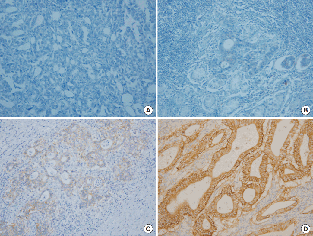
All sections were evaluated by a pathologist who did not have any clinical information on the patients. The pathologist scored slides as 0, 1+, 2+, or 3+ according to the gastric cancer HER-2 interpretive guidelines for immunohistochemistry. It was determined that 0 was no staining or membrane staining in < 10% of invasive tumor cells; 1+ was faint/barely perceptible membrane staining in ≥ 10% of invasive tumor cells–cells are only stained in part of their membranes; 2+ was weak to moderate complete or basolateral membrane staining in 10% of invasive tumor cells; and 3+ was moderate to strong complete or basolateral membrane staining in ≥10% of invasive tumor cells. In this study, intensity scores of 0, 1 + and 2+ were assumed to be negative, 3+ only was to be positive for overexpression. It is introduced in Fig. 1.
Statistical analysis
Statistical analysis was performed with the Statistical Package for Social Sciences PASW software ver. 18.0 (SPSS Inc., Chicago, IL, USA). The correlations between overexpression of HER-2 and clinicopathologic factors were evaluated using the Student’s t-test and chi-square test with Fisher’s exact test. Multivariate analyses were performed using logistic regression analysis on all variables found significant (> 20%) by univariate analyses. Kaplan-Meier curve analysis and Log rank test were used to evaluate overall survival and recurrence free survival probability. Significance was considered by two-sided test with a P-value of less than 0.05.
RESULTS
Clinicopathologic characteristics
Among 113 patients in this study, the range of patient age was 24–91 years and the median age was 65 years. HER-2 overexpression was evaluated in 16 (14.2%) patients. The clinicopathologic factors of 113 patients who underwent curative intent gastrectomy are arranged in Table 1.
Association with HER-2 overexpression and patient clinicopathologic factors
In this study’s results, positivity of HER-2 overexpression had statistically significant associations with tumor stage over IIIb (P= 0.036). Multivariated analysis showed the same result for factors that had significant association. Association of clinicopathologic factors and HER2 overexpression are arranged in Tables 2 and 3.
Association with HER2 overexpression and patient’s survival
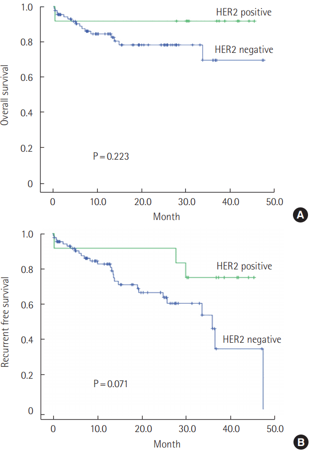
Among 113 patients, 5 patients (15.9%) died due to cancer, 13 patients were lost to follow-up, and 22 patients (19.5%) experienced recurrence. Because 13 patients were lost, the remaining 100 patients were included for survival analysis. The mean follow-up period was 17.4 months and the median follow-up period was 13.7 months (range, 0.4–47.6 months). In negative HER-2 overexpression, the 2-years overall survival (OS) rates were 78.1% and mean survival time was 37.5 months. In positive of HER-2 overexpression, it was 91.7% and 41.7 months. Between positive and negative of HER-2 overexpression, there was no significant difference for overall survival (P= 0.223) and recurrence free survival (P= 0.071). These are visualized in Fig. 2.
DISCUSSION
Through this study, we aimed to reveal the significant correlation between HER-2 overexpression in patients who underwent curative gastrectomy and their clinicopathologic factors. In our study, the positive rate of HER-2 overexpression was 14.2%, and this was similar to other studies [25].
HER-2 screening test indications have been established for breast cancer. The American Society of Clinical Oncology and the College of American Pathologists announced a clinical practice guideline for HER-2 testing for breast cancer [26]. According to this guideline, all patients with invasive breast cancer require their lesions be tested for HER-2 positivity; that test includes at least one of the two approved testing methods. And, if the cancer was metastatic, HER-2 screening tests would be done on the metastatic lesion. Especially, HER-2 positivity in breast cancer is related to a patient’s poor prognosis, and it is important to select the appropriate type of chemotherapy [9,14]. This guideline helps provide patients with options for HER-2 targeted therapy. The invention of HER-2 targeted therapy, Trastuzumab (Herceptin), induced steady improvement of treatment outcome on breast cancer [27,28].
Many studies have been done on HER-2 positivity in other cancers, like colon, lung, and stomach, as an increasing role of HER-2 positivity becomes more important in breast cancer. Especially, HER-2 positivity on gastric cancer, as in our study focus, was identified as a poor prognosis factor in gastric cancer patients, as with breast cancer [4,5,8,12,15-21]. However, up to now, research on the relevance of HER-2 positivity and gastric cancer is not sufficient to use in clinical practice. In Korea, HER-2 targeted therapy (Trastuzumab) is covered by our national health insurance system in HER-2 positive metastatic gastric and esophagogastric junctional cancer patients with no case of previous chemotherapy. Therefore evaluation of HER-2 status is important in Korea.
In other studies, HER-2 was evaluated as a prognostic factor of gastric cancer, though it is still not clarified. Son et al. [25] reported 15.1% of HER-2 positivity in gastric cancer underwent gastric surgery, and a significant correlation of HER-2 overexpression and histological grade and Lauren classification. Yet, this alone was not enough to evaluate prognostic values of HER-2 expression in gastric cancer. Oh et al. [11] reported HER-2 overexpression was associated with older age, differentiated histology, and intestinal type. Tanner et al. [8] reported an association between intestinal type and HER-2 overexpression, and they are associated with a poor outcome. In this study, we found that HER-2 has significant association with gastric cancer stage, as a specific setting of IIIb/IIIc. Additionally, this has no statistical meaning in other stage formats, but has meaning in revealing the association of HER-2 overexpression and gastric cancer stage. Especially, cancer stage is an important factor in patients’ poor survival outcome and prognosis, and sometimes related with operative plans, for instance, the extent of surgery [29,30]. Histologic findings had correlation with HER-2 overexpression (P= 0.055). It was similar with other studies wherein high grade differentiated gastric cancer had an association to positivity of HER-2 overexpression [16,18,19]. With a greater number of cases, this may be a significant result.
In fact, recent HER-2 overexpression has been estimated by chromogenic in situ hybridization, and fluorescence in situ hybridization. However, our study was only HER-2 overexpression in immunohistochemisty due to it being a retrospective study. In the HER-2 positivity interpretation, we assumed that 3+ only was to be positive for overexpression [20]. According to this criteria, it is believed that the number of positive groups is relatively low and the analysis associated with the characteristics has not found significant results. If the criteria was changed to over 2+ as positive for overexpression, it would be 40 cases of HER-2 positive [19]. In this interpretation, HER-2 overexpression is more often in high-grade differentiated gastric cancer (P< 0.001) [8,18].
In this study, we revealed that HER-2 overexpression was related to advanced stage. However, we did not find a correlation to patient survival because of the short-term follow-up period. If the period for analysis of survival rate were long enough, it would have a survival correlation with HER-2 overexpression. In this study, we only revealed an association between HER-2 and tumor stage. This study progressed in a single center. If there were more studies including multicenter and sufficient samples, it would be possible to define indications of HER-2 screening tests established in Korea. In the future, more effort is needed to discover specific biomarkers for gastric cancer and its prognostic factors.
In conclusion, we collected gastric cancer tissues from 113 patients with curative intent gastrectomy and evaluated the clinicopathologic factors related to HER-2 overexpression. Positive HER-2 overexpression rates were 14.2%, and patient tumor stage had association with positivity of HER-2 overexpression. Survival analysis of HER-2 overexpression has no significant difference.


