| Home | E-Submission | Sitemap | Contact Us |  
Korean Journal of Clinical Oncology > Article
Roh: Radiation segmentectomy for gastric leiomyosarcoma hepatic metastasis

ABSTRACT

Metastases to the liver can be found in various malignancies, most commonly originating from the colon, rectum, pancreas, stomach, esophagus, breast, lung, and melanoma. Surgical resection of liver metastasis is generally considered to be the definitive therapy fore cure. However, many patients are unable to undergo surgical resection due to medical comorbidities or multifocal extent of malignant disease affecting the liver. Among patients not eligible for surgery, other therapies exist for treatment in order to down stage the disease for surgical resection or for palliation. Radioembolization of hepatic metastases has shown to improve outcomes among patients with variety of malignancies including more common malignancies such as colorectal cancer. Yttrium-90 (Y-90) radioembolization has been successfully used in the management of hepatic metastases. A small series of metastatic sarcoma to the liver treated with radioembolization showed a promising response. We report a case of metastatic gastric leiomyosarcoma to the liver treated with Y-90 glass microspheres therapy using the radiation segmentectomy approach, previously described for hepatocellular carcinoma.

INTRODUCTION

Radioembolization of hepatic metastases improves outcomes in patients with a variety of malignancies [15]. Two commercially available microspheres, TheraSphere (BTG Corp., London, UK) and SIR-Spheres (Sirtex Medical Limited, North Sydney, Australia) contain β-emitting radioisotope yttrium-90 (Y-90) which results in local cellular destruction [6]. Radiation segmentectomy utilizes the concept of delivering high doses of radiation to ≤2 hepatic segments in order to ablate tumor [7]. This brief report describes a case of radiation segmentectomy for the treatment of metastatic gastric leiomyosarcoma to the liver.

CASE REPORT

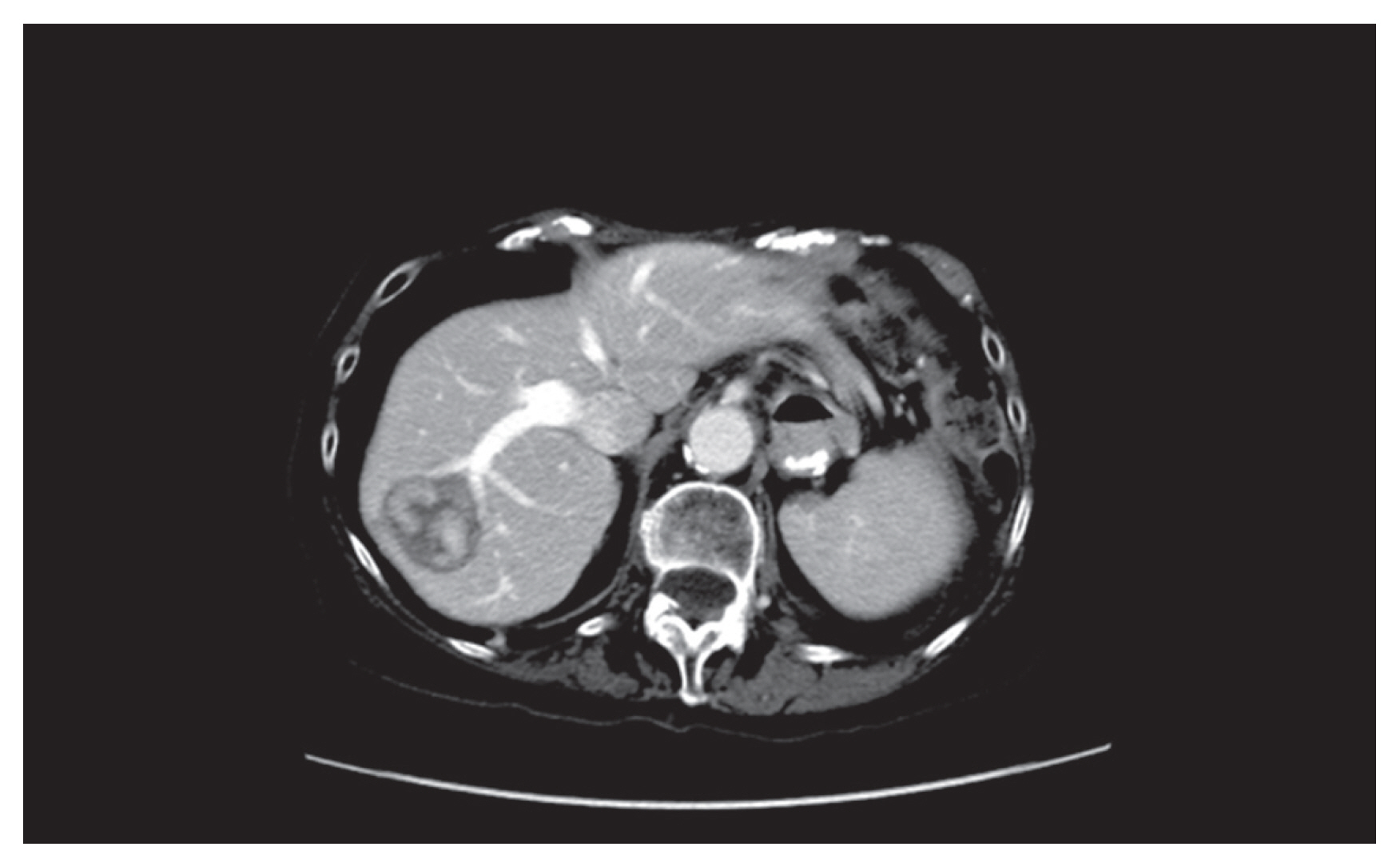
This case report was exempt from approval by Institutional Review Board of St. Luke’s University Health Network. An 86-yearold female with a history of high-grade gastric leiomyosarcoma developed metastasis to the liver (Fig. 1). The mass was biopsy proven; however, the pathologic analysis was unable to be obtained for this report. The patient had prior partial gastrectomy as well as chemotherapy in attempts to treat the malignancy. Despite the prior therapies, the patient was noted to have progression of disease with liver metastasis seen on a follow-up computed tomography. The patient was referred for locoregional therapy of her metastatic liver disease. The referring surgeon concluded that repeat surgery for resection of the hepatic metastasis would be very high risk.
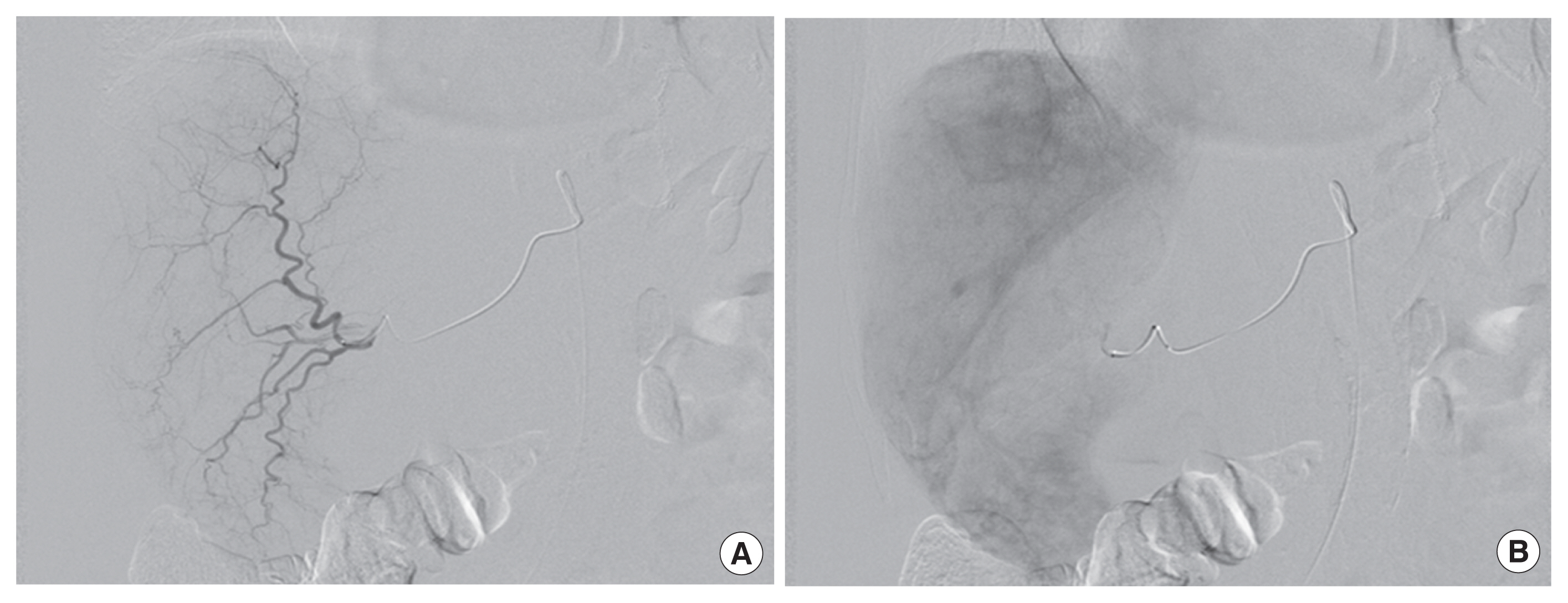
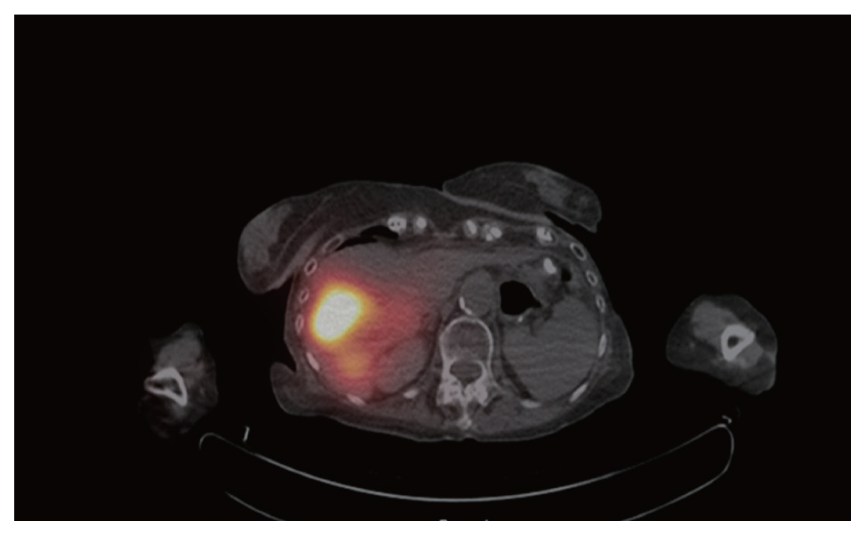
The right hepatic artery was selected using a 2.4-Fr Progreat microcatheter (Terumo, Tokyo, Japan). Angiography showed a hypervascular mass in segment 7 with arterial feeders arising from anterior and posterior divisions of the right hepatic artery (Fig. 2). The sub-lobar anterior division of the right hepatic artery supplying the segment 7 tumor was selectively cannulated and 73.7 mCi Y-90 glass microspheres (TheraSphere Corp.) were administered (Fig. 3A and B). This was followed by selective cannulation of the sub-lobar posterior division of the right hepatic artery and 75.8 mCi Y-90 glass microspheres were administered (Fig. 3C and D). Post-embolization Bremsstrahlung study shows good coverage of the metastatic lesion in segment 7 (Fig. 4).

DISCUSSION

Treatment of liver malignancies with radiation segmentectomy when the tumor is in a location unfavorable to ablation therapy has been described in the literature [2,7,8]. Radiation to ≤ 2 lobes is delivered with a high absorbed dose of 190 Gy with the intent to ablate the hepatic tumor [7]. The total radiation dose to be delivered is calculated with respect to the volume of the tumor that requires treatment.
Gastrointestinal stromal tumors (GISTs) are rare, comprising 0.1% to 3% of gastrointestinal tract malignancies [9]. Leiomyosarcomas, a subgroup of GISTs, are a rare collection of tumors with various origins such as stomach, uterus, kidney, retroperitoneum, and soft tissues [10]. Gastric leiomyosarcomas are extremely rare with no cases of metastatic gastric leiomyosarcomas to the liver reported in the literature.
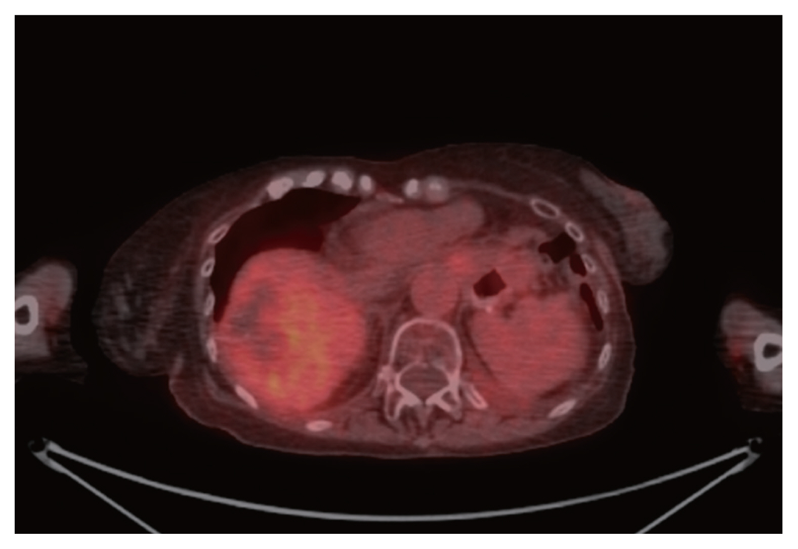
We report a case of gastric leiomyosarcoma metastatic to the liver treated with a high dose of Y-90 glass microspheres. The total radiation activity administered was 149.5 mCi with calculated absorbed dose to the metastatic liver lesion being greater than 190 Gy. The goal of this treatment is to induce necrosis within the entire volume of tumor treated. The patient responded to the treatment well and no significant uptake was seen within the tumor at 2 months imaging follow-up (Fig. 5). We recognize that a 2-month follow-up period is too short a period to assess for disease recurrence. We will be following up with the patient on a longer scale to determine if our therapy was curative.
The metastases treated in this report were hypovascular as can be seen in the angiographic images (Figs. 2, 3A and C). Obtaining computed tomography angiography prior to procedure can help to identify the specific vessels which need targeting, especially in the treatment of hypovascular metastases.
There have been prior reports of metastases to the liver treated with Y-90 therapy [11]. In a report by Sideras et al. [11], Y-90 resin microspheres were used to treat chemotherapy resistant nonseminomatous germ cell tumor (NSGCT) hepatic metastases. Although the concept of radiation segmentectomy did not exist at the publication time of the report, the concept used during the procedure, with less than two lobes of liver targeted resulting in superselective treatment, emulates radiation segmentectomy. The patient had excellent clinical and radiologic response. Further clinical trials are needed to determine the efficacy of radioembolization in management of chemotherapy resistant NSGCT metastases.
Metastatic leiomyosarcoma to the liver treated with Y-90 therapy has been described in the literature [12]. However, the abstract does not describe the organ of origin of the tumor. The abstract concluded that treating soft tissue sarcomas metastatic to the liver with Y-90 is safe, although no long-term follow-up was reported.
Our report shows that metastatic gastric leiomyosarcoma to the liver which chemotherapy had failed can be safely treated using Y-90 glass beads. It may be useful to treat other rare tumors metastatic to the liver of which systemic therapy had failed with Y-90 radioembolization.

ACKNOWLEDGMENTS

Author thanks Dr. Constantinos Sofocleous for the case report.

CONFLICT OF INTEREST

No potential conflict of interest relevant to this article was reported.

REFERENCES

1. Kennedy A, Cohn M, Coldwell DM, Drooz A, Ehrenwald E, Kaiser A, et al. Updated survival outcomes and analysis of long-term survivors from the MORE study on safety and efficacy of radioembolization in patients with unresectable colorectal cancer liver metastases. J Gastrointest Oncol 2017;8:614-24.
crossref pmid pmc
2. Boas FE, Bodei L, Sofocleous CT. Radioembolization of colorectal liver metastases: indications, technique, and outcomes. J Nucl Med 2017;58(Suppl 2):104S-111S.
crossref pmid
3. Bunck AC, Dos Santos DP, Chang DH, Reiser M, Pfister D, Bunck A, et al. Successful yttrium-90 microsphere radioembolization for hepatic metastases of prostate cancer. Case Rep Oncol 2017;10:627-33.
crossref pmid pmc
4. Jia Z, Jiang G, Zhu C, Wang K, Li S, Qin X. A systematic review of yttrium-90 radioembolization for unresectable liver metastases of melanoma. Eur J Radiol 2017;92:111-5.
crossref pmid
5. Gordon AC, Uddin OM, Riaz A, Salem R, Lewandowski RJ. Making the case: intra-arterial therapy for less common metastases. Semin Intervent Radiol 2017;34:132-9.
crossref pmid pmc
6. Venkatanarasimha N, Gogna A, Tong KT, Damodharan K, Chow PK, Lo RH, et al. Radioembolisation of hepatocellular carcinoma: a primer. Clin Radiol 2017;72:1002-13.
crossref pmid
7. Vouche M, Habib A, Ward TJ, Kim E, Kulik L, Ganger D, et al. Unresectable solitary hepatocellular carcinoma not amenable to radiofrequency ablation: multicenter radiology-pathology correlation and survival of radiation segmentectomy. Hepatology 2014;60:192-201.
crossref pmid
8. Riaz A, Gates VL, Atassi B, Lewandowski RJ, Mulcahy MF, Ryu RK, et al. Radiation segmentectomy: a novel approach to increase safety and efficacy of radioembolization. Int J Radiat Oncol Biol Phys 2011;79:163-71.
crossref pmid
9. Keung EZ, Raut CP. Management of gastrointestinal stromal tumors. Surg Clin North Am 2017;97:437-52.
crossref pmid
10. Rou WS, Ju JS, Kang SH, Moon HS, Sung JK, Lee BS, et al. A case of gastric leiomyosarcoma with multiple metastases. Korean J Gastroenterol 2015;65:112-7.
crossref pmid
11. Sideras PA, Sofocleous CT, Brody LA, Siegelbaum RH, Shah RP, Taskar NP. Superselective internal radiation with yttrium-90 microspheres in the management of a chemorefractory testicular liver metastasis. Cardiovasc Intervent Radiol 2012;35:426-9.
crossref pmid
12. In : Abstracts of the SIR 2015 Annual Scientific Meeting; February 28–March 5, 2015; Atlanta, GA. J Vasc Interv Radiol. 2015. 26(2 Suppl):p. S3-228.
pmid

Fig. 1
Pre-procedure computed tomography axial. A hypervascular metastatic lesion is present in segment 7 of liver.
kjco-14-2-142f1.jpg
Fig. 2
Intraoperative angiogram. (A) A microcatheter has selectively cannulated the right hepatic artery and its branches are clearly identified on this early arterial phase. (B) Delayed phase image shows a hypervascular mass corresponding to segment 7 lesion on prior computed tomography.
kjco-14-2-142f2.jpg
Fig. 3
Intraoperative angiogram and intraoperative cone beam computed tomography. Microcatheter has selectively cannulated the sub-lobar anterior (A) and posterior (C) division of right hepatic artery prior to delivery of yttrium-90 (Y-90) glass microspheres. (B, D) Dense contrast staining is present within the segment 7 hypervascular mass confirming successful targeting with Y-90 glass microspheres.
kjco-14-2-142f3.jpg
Fig. 4
Single photon emission computed tomography/computed tomography axial. Post-procedure yttrium-90 bremsstrahlung study shows good coverage of the segment 7 metastatic lesion.
kjco-14-2-142f4.jpg
Fig. 5
Two-month follow-up positron emission tomography-computed tomography axial. The segment 7 lesion shows hypometabolic activity indicating good response to radioembolization.
kjco-14-2-142f5.jpg
Editorial Office
101-3304 Brownstone Seoul, 464 Cheongpa-ro, Jung-gu, Seoul 04510, Korea
TEL : +82-2-393-2114   FAX : +82-2-393-1649   E-mail : ksco2004@daum.net

Copyright© Korean Society of Surgical Oncology.                Developed in M2PI
About |  Browse Articles |  Current Issue |  For Authors and Reviewers